Home » Without Label » Anatomy Rib Cage : Amazon Com Rib Cage Human Anatomy Antique Engraving Illustration Cool Wall Decor Art Print Poster 12x18 Posters Prints - This furrow isn't present in the 11th and 12th ribs.
Anatomy Rib Cage : Amazon Com Rib Cage Human Anatomy Antique Engraving Illustration Cool Wall Decor Art Print Poster 12x18 Posters Prints - This furrow isn't present in the 11th and 12th ribs.
Anatomy Rib Cage : Amazon Com Rib Cage Human Anatomy Antique Engraving Illustration Cool Wall Decor Art Print Poster 12x18 Posters Prints - This furrow isn't present in the 11th and 12th ribs.. The ribs are curved, flat bones which form the majority of the thoracic cage. Rib cage pain can be caused. Cavea thoracis, thoracic cage, rib cage, rib cage, thoracic cage, thoracic cage structure (body structure), thoracic cage structure, rib cage, nos. Rib bones are not classified as long bones.instead, anatomists classify the ribs as flat bones, and they are located within the axial skeleton.together with the sternum, thoracic vertebrae, and costal cartilages, the ribs form the thoracic cage, also called the bony thorax. They articulate with the vertebral column posteriorly, and terminate anteriorly as cartilage (known as costal cartilage).
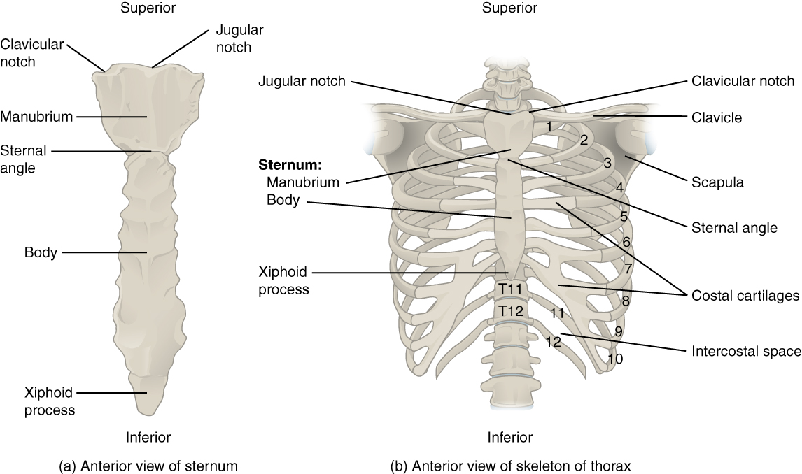
Elevates the ribs, increasing the thoracic volume. There are twelve pairs of ribs, all of which articulate with the vertebral column. Don't be fooled their long, curved shape! Rib bones are not classified as long bones.instead, anatomists classify the ribs as flat bones, and they are located within the axial skeleton.together with the sternum, thoracic vertebrae, and costal cartilages, the ribs form the thoracic cage, also called the bony thorax. In this image, you will find thoracic vertebrum, costochondral joint, costal cartilage, costal margin, costal arch, thoracic vertebrum, xiphoid process, xiphisternal joint, body, manubrial sternal joint, manubrium, the sternal notch in it.
File 721 Rib Cage Jpg Wikimedia Commons from upload.wikimedia.org The rib cage is the arrangement of ribs attached to the vertebral column and sternum in the thorax of most vertebrates, that encloses and protects the vital organs such as the heart, lungs and great vessels. Ten of the twelve ribs connect to strips of hyaline cartilage on the anterior side of the body. An enlarged or ruptured spleen can cause sudden or chronic pain under the left rib cage that ends up migrating towards the back and/or shoulders. The thoracic cage (rib cage) is the skeletal framework of the thoracic wall, which encloses the thoracic cavity. Anatomy of the rib cage. As part of the bony thorax, the ribs protect the internal thoracic organs. Contributing to their role in protecting the internal thoracic organs. Rib cage, in vertebrate anatomy, basketlike skeletal structure that forms the chest, or thorax, and is made up of the ribs and their corresponding attachments to the sternum (breastbone) and the vertebral column.
The rib cage, which forms the chest wall, is an important volume.
They are extremely light, but highly resilient; Click the image to watch the anatomy of the rib cage video. Human muscles · april 17, 2020. The upper edge is round and the lower sharp. Elevates the ribs, increasing the thoracic volume. Womens body parts stomach 4 photos of the womens body parts stomach body diagram stomach, body parts digestive system, body parts in stomach area, body parts liver, body parts spleen, human body parts stomach, woman body organs, woman body parts found, stomach, body diagram stomach, body parts digestive system, body. However, only seven have a direct articulation with the sternum. #proko #art #anatomy #ribs #ribcage #humananatomy #tutorial. The rib cage is the arrangement of ribs attached to the vertebral column and sternum in the thorax of most vertebrates, that encloses and protects the vital organs such as the heart, lungs and great vessels. In this image, you will find thoracic vertebrum, costochondral joint, costal cartilage, costal margin, costal arch, thoracic vertebrum, xiphoid process, xiphisternal joint, body, manubrial sternal joint, manubrium, the sternal notch in it. Anatomy the rib cage is a bony structure found in the chest (thoracic cavity). Shaped somewhat like a cone, it is created by the individual ribs connecting to the spine above and to the sternum below. It consists of the 12 pairs of ribs with their costal cartilages and the sternum (figure 6.38).
There are twelve pairs of ribs, all of which articulate with the vertebral column. Related posts of rib cage diagram with organs womens body parts stomach. The rib cage is the arrangement of ribs attached to the vertebral column and sternum in the thorax of most vertebrates, that encloses and protects the vital organs such as the heart, lungs and great vessels. It is made up of 12 pairs of ribs. The rib cage consists of 24 ribs, 12 on either side, and it shields the organs of the chest, including the heart and the lungs, from damage.
The Thoracic Cage The Rib Cage Youtube from i.ytimg.com The cartilage strips are called costal cartilage (costal is the anatomical adjective that refers to the rib) and connect on their other end to the sternum. Rib cage, in vertebrate anatomy, basketlike skeletal structure that forms the chest, or thorax, and is made up of the ribs and their corresponding attachments to the sternum (breastbone) and the vertebral column. The thoracic cage consists of the 12 thoracic vertebrae, the associated intervertebral discs, 12 pairs of ribs with their costal cartilages, and the sternum. The thoracic cage protects the heart and lungs. Rib cage pain may be sharp, dull, or achy and felt at or below the chest or above the navel on either side. It consists of the 12 pairs of ribs with their costal cartilages and the sternum (figure 6.38). The rib cage, which forms the chest wall, is an important volume. Anatomy the rib cage is a bony structure found in the chest (thoracic cavity).
It is made up of 12 pairs of ribs.
Shaped somewhat like a cone, it is created by the individual ribs connecting to the spine above and to the sternum below. Derived from the nih umls ( unified medical language. The ribs are a set of twelve paired bones which form the protective 'cage' of the thorax. Elevates the ribs, increasing the thoracic volume. The thoracic cage (rib cage) is the skeletal framework of the thoracic wall, which encloses the thoracic cavity. The thoracic cage protects the heart and lungs. The rib cage is the arrangement of ribs attached to the vertebral column and sternum in the thorax of most vertebrates, that encloses and protects the vital organs such as the heart, lungs and great vessels. The rib cage, which forms the chest wall, is an important volume. The rib cage consists of 24 ribs, 12 on either side, and it shields the organs of the chest, including the heart and the lungs, from damage. The primary causes of pain under the left rib cage. The average skeleton contains 24 individual ribs, formed in 12. Originate at the lower border of the rib, inserting into the superior border of the rib below. Human muscles · april 17, 2020.
The cartilage strips are called costal cartilage (costal is the anatomical adjective that refers to the rib) and connect on their other end to the sternum. The rib below that is rib 2, and it connects to the t2 thoracic vertebra, and so on. Check out our anatomy rib cage selection for the very best in unique or custom, handmade pieces from our shops. The thoracic cage (rib cage) is the skeletal framework of the thoracic wall, which encloses the thoracic cavity. It may occur after an obvious injury or without explanation.
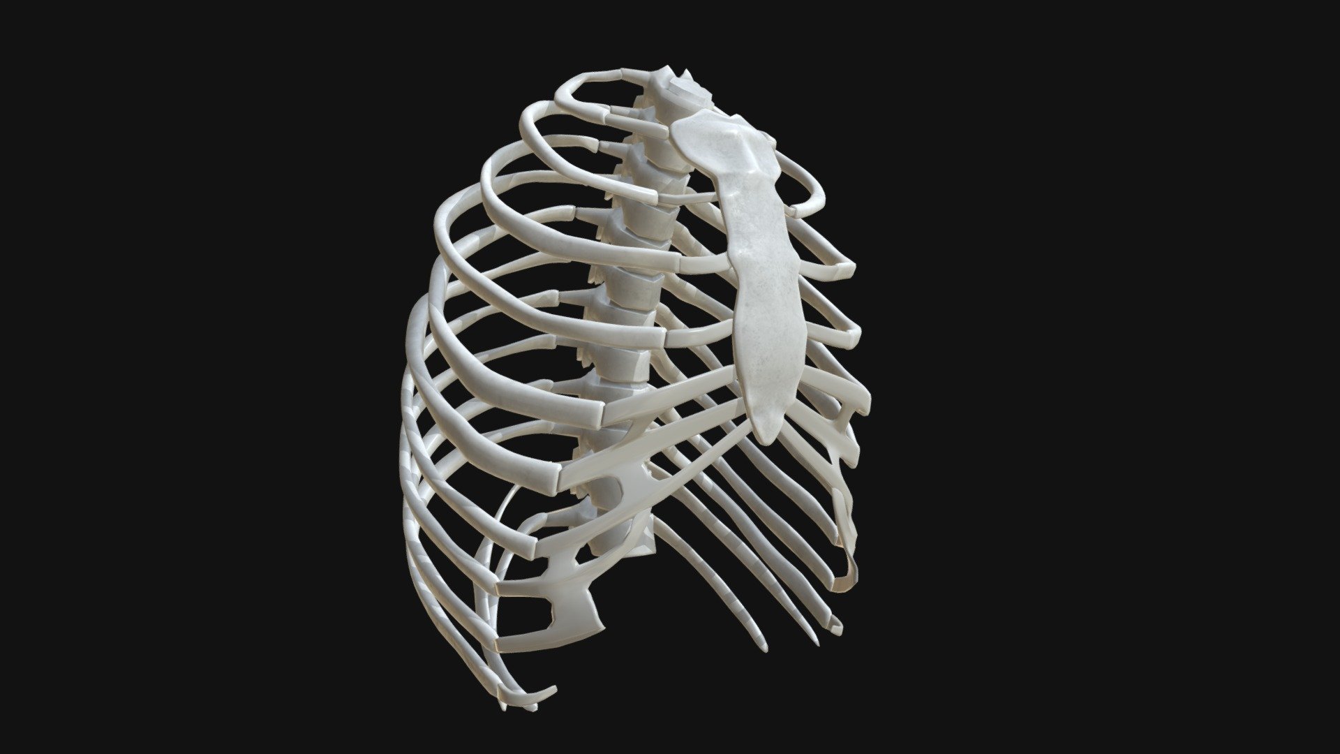
Anatomy Human Rib Cage Buy Royalty Free 3d Model By Francescomilanese Francescomilanese 0f1aa77 from media.sketchfab.com Rib cage pain may be sharp, dull, or achy and felt at or below the chest or above the navel on either side. Anatomy the rib cage is a bony structure found in the chest (thoracic cavity). Rib cage, in vertebrate anatomy, basketlike skeletal structure that forms the chest, or thorax, and is made up of the ribs and their corresponding attachments to the sternum (breastbone) and the vertebral column. Cavidad costal, estructura de la caja torácica (estructura corporal), estructura de la caja torácica, jaula costal. Rib cage pain can be caused. Contributing to their role in protecting the internal thoracic organs. However, only seven have a direct articulation with the sternum. The rib below that is rib 2, and it connects to the t2 thoracic vertebra, and so on.
Derived from the nih umls ( unified medical language.
Shaped somewhat like a cone, it is created by the individual ribs connecting to the spine above and to the sternum below. Derived from the nih umls ( unified medical language. There are twelve pairs of ribs, all of which articulate with the vertebral column. Ten of the twelve ribs connect to strips of hyaline cartilage on the anterior side of the body. Related posts of rib cage diagram with organs womens body parts stomach. Don't be fooled their long, curved shape! The rib below that is rib 2, and it connects to the t2 thoracic vertebra, and so on. The ribs are a set of twelve paired bones which form the protective 'cage' of the thorax. However, only seven have a direct articulation with the sternum. #proko #art #anatomy #ribs #ribcage #humananatomy #tutorial. The average skeleton contains 24 individual ribs, formed in 12. Rib bones are not classified as long bones.instead, anatomists classify the ribs as flat bones, and they are located within the axial skeleton.together with the sternum, thoracic vertebrae, and costal cartilages, the ribs form the thoracic cage, also called the bony thorax. The upper edge is round and the lower sharp.